BREIT
Brain Stroke Reconstruction using Multi-Frequency 3D EIT
BREIT: A Framework for Brain Stroke Reconstruction using Multi-Frequency 3D EIT
1 ETIS (UMR 8051), CY Cergy Paris University, ENSEA, CNRS · 2 AGM (UMR 8088), CY Cergy Paris University, CNRS
3 Paris-Saclay University, CentraleSupélec, Inria, CVN
* Corresponding author
The MICCAI 2026 proceedings paper represents the peer-reviewed version of record (link forthcoming). The earlier arXiv preprint differs from the final camera-ready version.
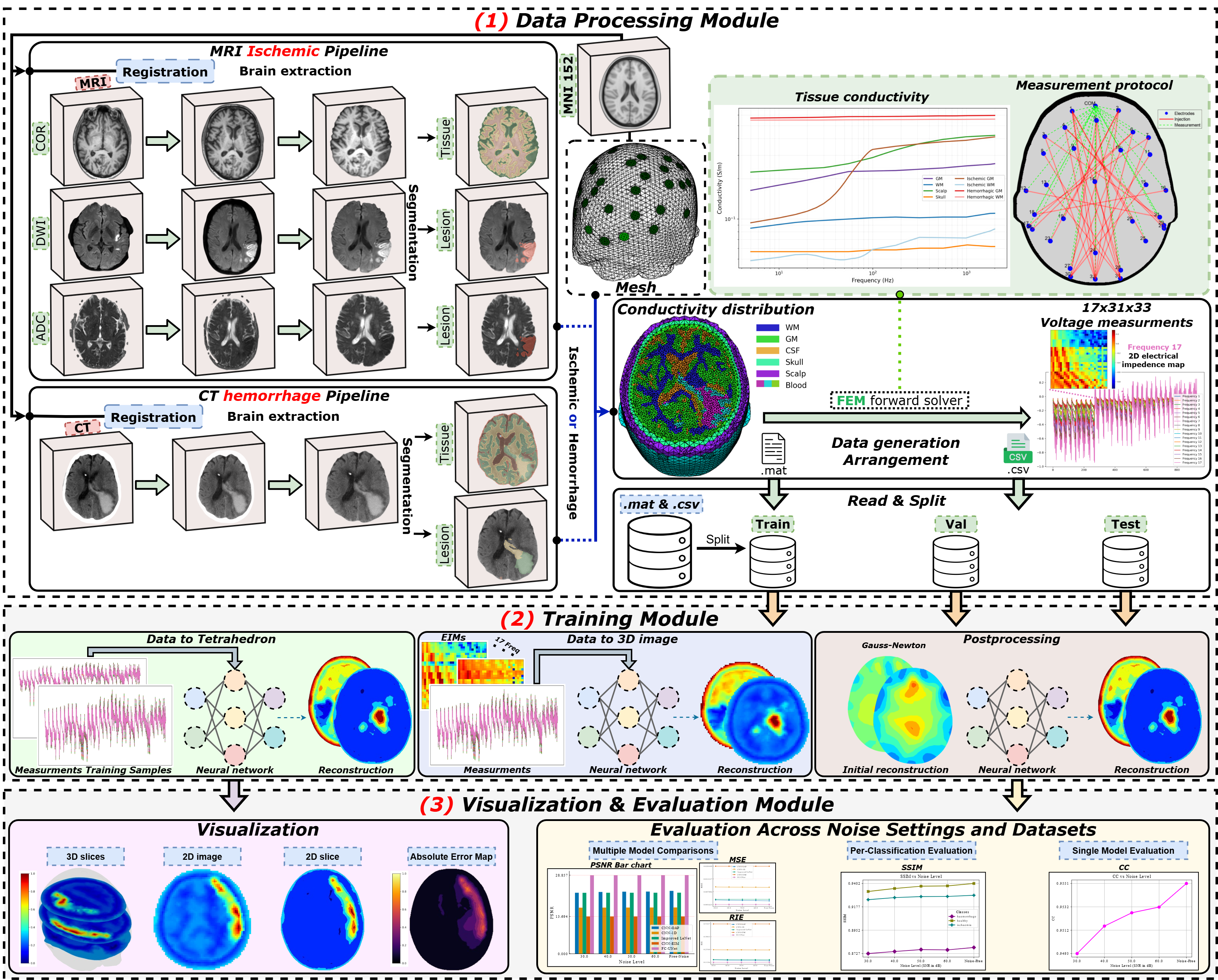
The BREIT framework. A modular pipeline for 3D MF-EIT stroke reconstruction: neuroimaging-to-admittivity ground-truth generation, a Python 3D CEM forward solver, and a 3D D-bar implementation supporting non-uniform electrode layouts.
Abstract
Multi-Frequency Electrical Impedance Tomography (MF-EIT) is a non-invasive, low-cost modality that reconstructs electrical property distributions from boundary voltages. For stroke imaging, progress in 3D deep-learning reconstruction is limited by the lack of large-scale datasets with paired ground-truth volumes and by non-standardized pipelines for data generation, simulation, and evaluation. We introduce BREIT, a modular framework for 3D MF-EIT stroke reconstruction providing: (i) a neuroimaging-to-EIT pipeline that converts CT/MRI into frequency-dependent GT admittivity volumes; (ii) a self-contained Python 3D Complete Electrode Model (CEM) forward solver for simulating MF-EIT voltages; and (iii) a 3D D-bar implementation supporting non-uniform electrode layouts. Building on BREIT, we propose dFNO-bar, which integrates Fourier Neural Operators into D-bar by learning a mapping from scattering data t(ξ) to conductivity σ(x)=ℜ{γ}. We evaluate dFNO-bar against D-bar, Deep D-bar, and Gauss–Newton reconstructions on UCLH-matched synthetic data, and observe higher brain SSIM with comparable CC across noise settings.
Highlights
Key Contributions
BREIT unifies radiology-based admittivity generation, realistic MF-EIT simulation, and DL benchmarking under consistent configurations.
Multimodal Data Generation
CT/MRI-based tissue and lesion mapping to 3D frequency-dependent admittivity volumes, paired with simulated MF-EIT voltages across 17 frequencies.
3D CEM + D-bar Tooling
A Python implementation of 3D D-bar components including texp and t0 approximations for non-uniform electrode layouts with Voronoi weighting.
dFNO-bar Reconstruction
A 3D MF-EIT reconstruction model that couples Fourier Neural Operator learning with D-bar structure for improved stroke imaging.
The Method
BREIT Pipeline
A four-stage workflow for generating clinically plausible MF-EIT data and simulations: patient-specific data curation, imaging-driven ground-truth approximation, physics-consistent augmentation, and multi-frequency forward-model voltage synthesis.
Clinical Imaging to EIT-Ready Volumes
CT and MRI studies are collected, aligned, and converted into consistent 3D head representations with tissue-level labels and lesion localization used for subsequent electrical property modeling.
Neuroimaging to Frequency-Dependent Admittivity
Tissue compartments are mapped to conductivity and permittivity priors across frequency, producing voxel-wise admittivity targets that serve as reconstruction ground truth.
Diversity and Robustness Enrichment
Subject variability is expanded with controlled perturbations of lesion extent, position, and tissue-property ranges so training covers realistic anatomical and pathological uncertainty.
3D CEM Multi-Frequency Voltage Synthesis
A Python 3D Complete Electrode Model solver simulates boundary voltages for non-uniform electrode layouts over all acquisition frequencies, yielding paired inputs for reconstruction learning and evaluation.
Validation
Forward Solver Agreement with Clinical MF-EIT
We validate simulation realism by comparing forward-simulated voltages to corresponding clinical MF-EIT measurements in UCLH for all subjects with matched imaging (12 patients, 14 sessions).
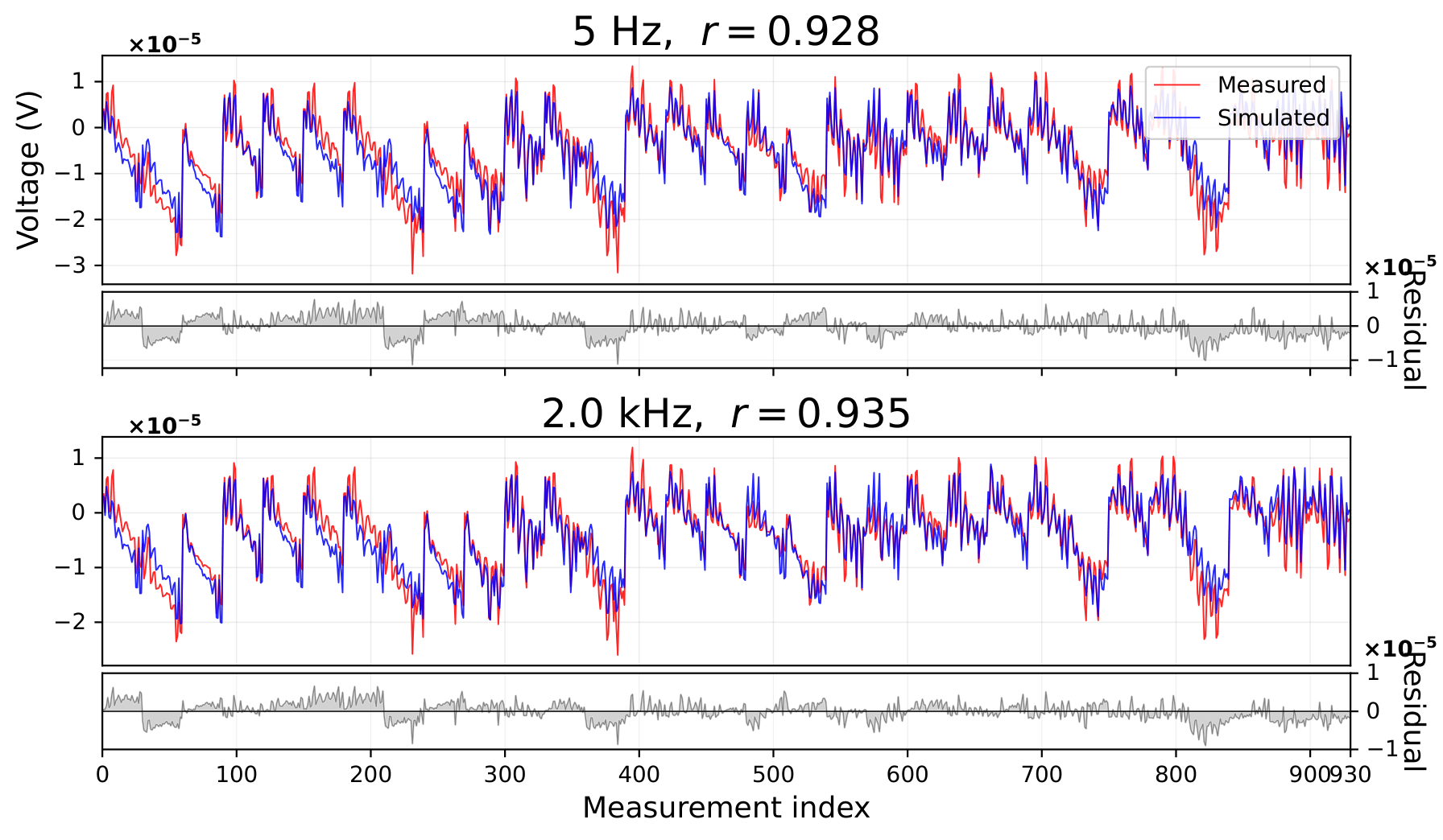
Forward-solver voltage agreement with clinical MF-EIT. Overlays for pat-01 at 5 Hz and 2 kHz after per-frequency affine alignment, showing strong correlation (r = 0.928 at 5 Hz, r = 0.935 at 2 kHz).
Reconstruction
dFNO-bar Architecture
dFNO-bar integrates Fourier Neural Operators into a D-bar reconstruction pipeline, learning a mapping from multi-truncation scattering data to spatial-domain conductivity.
Classical D-bar: Stability Through Truncation
Conventional D-bar reconstruction improves robustness by truncating high-frequency scattering components t(ξ), which are generally more sensitive to measurement noise. While this regularization strategy stabilizes the inverse problem and reduces artifacts, it may also remove informative structural content that contributes to accurate conductivity recovery, particularly near fine anatomical boundaries.
Learning Information Across Truncation Levels
Rather than enforcing a single truncation radius, dFNO-bar provides multiple complex scattering truncations Tξ as parallel input channels. The FNO learns which higher-frequency components should be preserved, attenuated, or denoised, yielding a data-driven balance between stability and information retention. This design naturally complements the D-bar recovery stage that maps scattering-domain data into conductivity estimates.
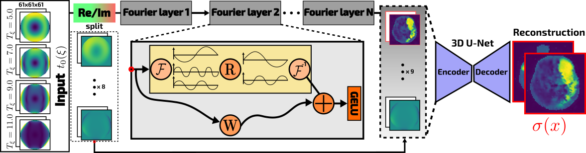
Proposed dFNO-bar architecture. A FNO maps the input in Fourier space (split real/imag from 4 truncation radii) through stacked Fourier blocks, followed by a lightweight 3D U-Net decoder to produce the spatial-domain output conductivity σ(x).
Results
Quantitative & Visual Comparison
dFNO-bar achieves consistently higher brain SSIM and comparable CC to Deep D-bar across noise settings, with fewer parameters and lower memory usage. Below, SSIM and CC are reported as averages over noiseless, 50 dB, and 40 dB settings.






Animated qualitative comparison. Two pathology cases are shown with matched frame-wise reconstructions from D-bar, Deep D-bar, and dFNO-bar against ground truth.
Citation
BibTeX
If you find BREIT useful, please cite the paper.
@inproceedings{abdelmoumene2026breit,
  title={BREIT: A Framework for Brain Stroke Reconstruction using Multi-Frequency 3D EIT},
  author={Abdelmoumene, Djahid and Ayad, Ishak and Nguyen, Ma{\"i} K. and Daveau, Christian},
  booktitle={International Conference on Medical Image Computing and Computer-Assisted Intervention (MICCAI)},
  year={2026}
}